Enamel hypoplasia
Insufficient enamel formation or mineralization during tooth development, which may cause abnormal color, surface defects, sensitivity, and increased caries risk.
Common Symptoms
Recognizing Enamel hypoplasia
Focus on the most useful decision cues first: common symptoms, the patients or situations that usually prompt review, and any signs that need faster assessment.
Common Symptoms
Signs patients often notice before evaluation
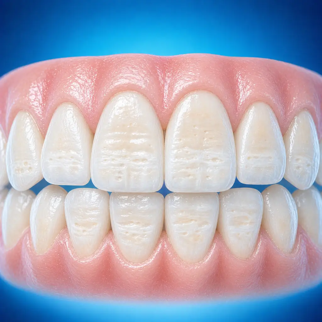
Mild cases may show only chalky white or yellow-brown patches or altered translucency, usually without obvious pain
Moderate to severe cases may show pinpoint, pit-and-groove, or band-like enamel defects; the tooth surface is rough and plaque retention is easy
When dentin is exposed, sensitivity to cold, heat, sour, or sweet stimuli may occur, especially when children eat or brush
Defective teeth are more prone to caries, wear, thinning of incisal edges, or cusp defects, often symmetrically or across multiple teeth
When to Seek Evaluation
Typical patients and situations that warrant review
Common in children and adolescents with permanent dentition, because permanent enamel mineralization takes a long time, from birth to age 6-7, and is easily affected by systemic factors
Tooth structure defects or abnormal enamel color
Rough, dull tooth surfaces with symmetrical distribution
Enamel fractures easily or shows obvious wear
Tooth sensitivity affects eating
Treatment Approaches
Treatment Directions for Enamel hypoplasia
Mild cases may be treated with remineralization or bleaching techniques to improve appearance
Moderate defects, such as pits and grooves, may be restored with composite resin to restore tooth shape and color
Severe defects, such as large areas of enamel loss or dentin exposure, require porcelain veneers or full crowns
For sensitivity, desensitizing treatment may be performed first
Children may first receive conservative treatment, such as fluoride application and resin fillings, followed by definitive restoration after adulthood
For severe generalized full-mouth defects, full-mouth occlusal rehabilitation may be considered
What usually shapes the treatment plan
Clinical Assessment
Key Assessments for Enamel hypoplasia
These are the main areas doctors usually review first. If you already have relevant test or imaging reports, bring them to speed up the assessment. They are helpful but not required, and the same workup can also be completed in China.
Severity of enamel defects
Whether dentin is exposed
Secondary caries
Tooth sensitivity
Before You Travel
How to Prepare
Maintain oral cleanliness
Planning Notes
Pre-Assessment Required
An oral specialist should perform an intraoral examination and, as appropriate, periodontal probing, pulp vitality testing, periapical radiographs, panoramic radiographs, or CBCT before determining the treatment plan. Key checks include visual assessment of enamel surface shape, color, defect features, and distribution; probing to assess defect depth and dentin exposure; percussion to assess pulp symptoms; and radiographs to evaluate root development, defect depth, secondary caries, and periapical disease. Bring oral examination and radiograph records if available.
Remote Pre-Assessment
Intraoral photos, the course of pain/swelling, previous dental records, and imaging can be submitted remotely for preliminary triage, urgency assessment, and an estimated treatment direction. Final diagnosis still requires in-person intraoral examination and necessary imaging.
Multidisciplinary Assessment
Medical History Important
Previous dental treatment history, imaging, allergy history, anticoagulant/bisphosphonate use, diabetes, and immune-related diseases can affect diagnosis, anesthesia, bleeding and infection risk, and treatment selection.
Ready to Explore Treatment for Enamel hypoplasia?
Let Carevia help you connect with the right specialists, compare hospitals, and plan your medical trip to China.