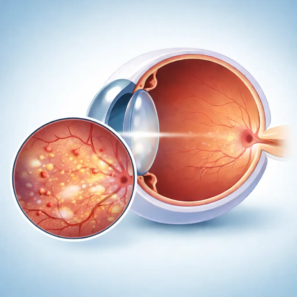
Diabetic Retinopathy
One of the most common microvascular complications of diabetes, causing retinal hemorrhage, edema, and neovascularization that can lead to blindness.
Common Symptoms
Recognizing Diabetic Retinopathy
Focus on the most useful decision cues first: common symptoms, the patients or situations that usually prompt review, and any signs that need faster assessment.
Common Symptoms
Signs patients often notice before evaluation
Usually asymptomatic in early stages
Gradual vision decline
Floating dark spots (vitreous hemorrhage)
Distorted vision (macular edema)
Sudden vision loss (significant vitreous hemorrhage or retinal detachment)
When to Seek Evaluation
Typical patients and situations that warrant review
Type 1 and Type 2 diabetes patients
Long-duration diabetes (>60% incidence at 10+ years)
Those with poor glycemic control
Concurrent hypertension and dyslipidemia
Gestational diabetes patients
First fundus screening should occur soon after diabetes diagnosis
Any change in vision
Diabetes duration over 5 years without prior fundus exam
Known DR requiring regular follow-up
Urgent Assessment
Sudden onset of numerous floaters or acute vision loss may indicate vitreous hemorrhage or tractional retinal detachment. Seek emergency care immediately.
Treatment Approaches
Treatment Directions for Diabetic Retinopathy
Blood glucose, blood pressure, and lipid control as foundation
Anti-VEGF injection (for DME and proliferative DR)
Retinal laser photocoagulation (treat ischemic areas, prevent neovascularization)
Vitrectomy (for severe vitreous hemorrhage and tractional retinal detachment)
What usually shapes the treatment plan
Clinical Assessment
Key Assessments for Diabetic Retinopathy
These are the main areas doctors usually review first. If you already have relevant test or imaging reports, bring them to speed up the assessment. They are helpful but not required, and the same workup can also be completed in China.
Dilated fundus examination
OCT (assess macular edema)
OCTA (evaluate retinal microvascular abnormalities)
Fluorescein angiography (assess ischemic areas and neovascularization)
Glycated hemoglobin (HbA1c) level
Before You Travel
How to Prepare
Bring recent HbA1c and blood glucose monitoring records
Bring previous fundus examination reports and angiography images
Prepare diabetes medication regimen details
Know recent blood pressure and lipid control status
Planning Notes
Pre-Assessment Required
Comprehensive fundus evaluation and systemic metabolic assessment required, including dilated fundus photography, OCT, FFA, HbA1c, blood pressure, and lipid levels to determine DR staging and treatment plan.
Remote Pre-Assessment
Fundus photographs, OCT reports, and HbA1c results can be submitted remotely for preliminary DR staging assessment and treatment recommendations. Angiography and surgical evaluation require on-site visits.
Multidisciplinary Assessment
Severe DR often requires ophthalmology-endocrinology collaboration to optimize glycemic management alongside eye treatment. Complex cases may also involve nephrology.
Medical History Important
Diabetes duration, glycemic control history, hypoglycemic regimen, and comorbidities (nephropathy, cardiovascular disease) all directly influence DR treatment decisions and prognosis.
Ready to Explore Treatment for Diabetic Retinopathy?
Let Carevia help you connect with the right specialists, compare hospitals, and plan your medical trip to China.