Dental fluorosis
Abnormal enamel color and structure caused by excessive fluoride intake during tooth development. It mainly affects appearance, and severe cases require restorative treatment.
Common Symptoms
Recognizing Dental fluorosis
Focus on the most useful decision cues first: common symptoms, the patients or situations that usually prompt review, and any signs that need faster assessment.
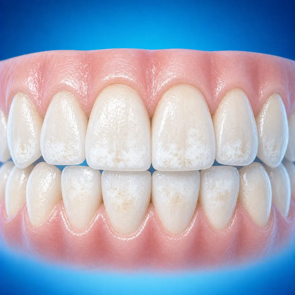
Common Symptoms
Signs patients often notice before evaluation
Teeth erupting during the same period may show chalky to brown patches on the enamel, and severe cases also have actual enamel defects. Clinically, it is often classified by severity as chalky, stained, or defective type
Because of the placental barrier, fluorosis mostly affects permanent teeth and rarely affects primary teeth; when primary teeth are affected, the degree is usually mild. If fluoride intake is excessive beyond the placental barrier's capacity, primary teeth may also be affected irregularly
Resistant to acid but not to wear
Severe chronic fluoride poisoning may cause bone overgrowth and calcification of periosteal ligaments. Acute poisoning may cause nausea, vomiting, and diarrhea
When to Seek Evaluation
Typical patients and situations that warrant review
Common in children and adolescents during permanent tooth eruption
People who lived long term before age 6-7 in endemic areas with high fluoride drinking water; even if they later move elsewhere, later-erupting permanent teeth may still be affected
Chalky opaque areas on the tooth surface covering more than one quarter of the surface or even the whole surface, with loss of luster
Yellow, yellow-brown, or dark brown tooth staining that cannot be scraped off
Shallow pits or concave defects on the tooth surface, with a shallow honeycomb-like appearance
Abnormal tooth color or rough, uneven enamel surface seen immediately after permanent tooth eruption
Treatment Approaches
Treatment Directions for Dental fluorosis
Mild dental fluorosis, chalky type, may be treated with bleaching and remineralization to remove surface stains and improve appearance
Moderate dental fluorosis, stained type, may be treated with resin infiltration, bleaching, or porcelain veneers to mask or improve tooth color
Severe dental fluorosis, defective type, requires composite resin restoration, porcelain veneers, or full crowns to restore tooth shape
What usually shapes the treatment plan
Clinical Assessment
Key Assessments for Dental fluorosis
These are the main areas doctors usually review first. If you already have relevant test or imaging reports, bring them to speed up the assessment. They are helpful but not required, and the same workup can also be completed in China.
Severity of dental fluorosis, mild, moderate, or severe, and lesion type, chalky, stained, or defective
Range and distribution of affected teeth, including whether involvement is symmetrical and affects multiple teeth
Whether tooth structure defects or secondary caries are present
Patient age and esthetic needs
Whether systemic fluoride toxicity, such as skeletal fluorosis, is present
History of living in a high-fluoride area
Before You Travel
How to Prepare
Maintain oral cleanliness
Planning Notes
Pre-Assessment Required
An oral specialist should perform an intraoral examination and, as appropriate, periodontal probing, pulp vitality testing, periapical radiographs, panoramic radiographs, or CBCT before determining the treatment plan. Key checks include visual assessment of tooth color, luster, and defects; probing to assess enamel hardness and defect depth; percussion to assess periapical problems; and radiographs to evaluate whether other tooth or pulp diseases are present. Bring imaging and specialist oral examination records if available.
Remote Pre-Assessment
Intraoral photos, the course of pain/swelling, previous dental records, and imaging can be submitted remotely for preliminary triage, urgency assessment, and an estimated treatment direction. Final diagnosis still requires in-person intraoral examination and necessary imaging.
Multidisciplinary Assessment
Medical History Important
Previous dental treatment history, imaging, allergy history, anticoagulant/bisphosphonate use, diabetes, and immune-related diseases can affect diagnosis, anesthesia, bleeding and infection risk, and treatment selection.
Ready to Explore Treatment for Dental fluorosis?
Let Carevia help you connect with the right specialists, compare hospitals, and plan your medical trip to China.