SMILE Laser Surgery (Small Incision Lenticule Extraction)
A flapless all-femtosecond laser procedure that removes a precisely shaped corneal lenticule through a ~2 mm micro-incision to correct myopia and astigmatism, with minimal dry eye and rapid recovery.
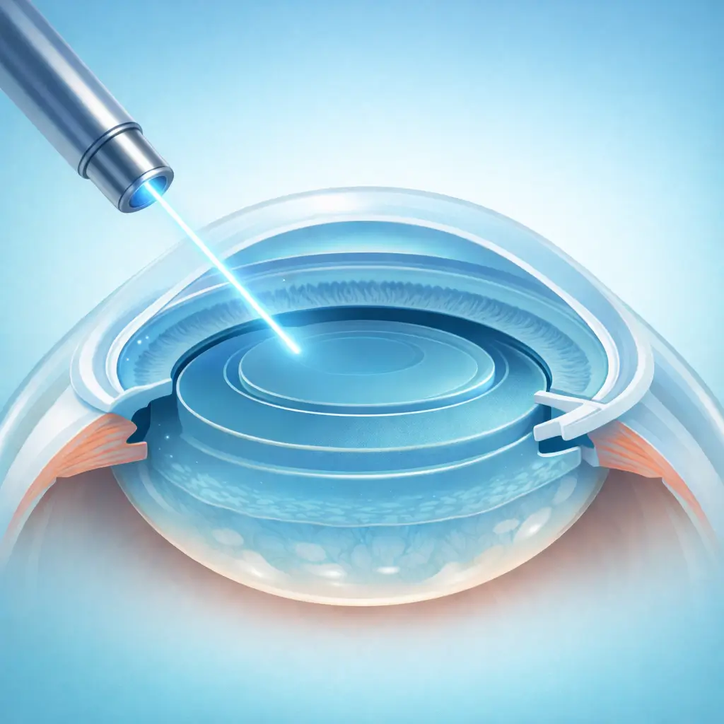
SMILE (Small Incision Lenticule Extraction) is one of the most advanced laser vision correction procedures available. Unlike LASIK, no corneal flap is created. Instead, a femtosecond laser precisely carves a disc-shaped lenticule within the corneal stroma, which is then extracted through a ~2 mm micro-incision to reshape the cornea and correct myopia and astigmatism. The Zeiss VisuMax femtosecond system used for SMILE dominates global procedure volumes, and major refractive centers in China's first-tier cities perform tens of thousands of SMILE procedures annually, accumulating extraordinary clinical experience. SMILE's principal advantages include preservation of the anterior stromal nerve plexus and Bowman's layer, which substantially reduces postoperative dry eye compared with LASIK, and eliminates flap-related complications, making it well-suited for patients in contact sports. Corneal biomechanical stability is also superior to LASIK. However, SMILE currently requires the Zeiss VisuMax platform, and astigmatism correction is limited (typically ≤3.00 D); patients outside this range may be better served by LASIK or TransPRK. Contact lens wear must be discontinued before the preoperative examination: soft lenses for at least 1 week, rigid gas-permeable (RGP) lenses for at least 1 month, and orthokeratology lenses for at least 3 months.
Quick Reference
Treatment
10 mins – 20 mins
Observation
1 hours – 2 hours
Est. Cost
$2,700 – $5,600
Department
Ophthalmology
Who Is This For
Is SMILE Laser Surgery (Small Incision Lenticule Extraction) Right for You?
Good Candidates
- Age 18-45 with stable myopia for ≥2 years (change ≤0.50 D/year)
- Myopia between -1.00 D and -10.00 D with astigmatism ≤3.00 D
- Adequate corneal thickness (central pachymetry usually ≥480 μm; residual stromal bed ≥250 μm post-treatment)
- Normal corneal topography with no keratoconus signs
- Mild or absent dry eye with essentially normal tear function
May Not Be Suitable
- Keratoconus or suspicious corneal topography patterns
- Thin cornea (<480 μm) or insufficient residual stromal bed after planned correction
- Progressive myopia within the past 2 years (>0.50 D/year)
- Active dry eye disease, severe meibomian gland dysfunction, or Sjögren's syndrome
- Systemic autoimmune diseases (e.g., rheumatoid arthritis, systemic lupus erythematosus)
Step-by-Step Process
How SMILE Laser Surgery (Small Incision Lenticule Extraction) Works
Preparation and Topical Anesthesia
The patient lies supine. Topical anesthetic drops are instilled bilaterally. The surgical eye is sterilized and draped; a lid speculum holds the eye open. The center is aligned with the laser treatment zone.
Femtosecond Laser Lenticule Creation
A suction cup docks with the eye. The Zeiss VisuMax femtosecond laser (500 kHz repetition rate) sequentially cuts the posterior and anterior lenticule surfaces within the stromal bed, shaping a disc corresponding to the desired refractive correction and creating a ~2 mm surface incision. The entire laser application takes approximately 20-30 seconds.
Interface Dissection
A fine spatula is inserted through the micro-incision to gently dissect the lenticule's upper and lower interfaces from the surrounding stroma, ensuring the lenticule is fully mobile.
Lenticule Extraction
Micro-forceps grasp and extract the free lenticule through the micro-incision. The lenticule is inspected to confirm it is intact and complete.
Irrigation and Discharge
The interface is gently irrigated with balanced salt solution. Antibiotic drops are instilled. No sutures or eye patch are needed. The patient rests in the recovery area for 60-120 minutes and is then discharged.
Both eyes are treated in a single visit on the same day. Follow-up visits are scheduled at day 1, week 1, months 1, 3, 6, and 12, then annually.
Cost Information
Cost Estimate for SMILE Laser Surgery (Small Incision Lenticule Extraction)
Estimated Price Range
$2,700 – $5,600
What's Included
Public tier-3A International Medical Department: approximately ¥18,000-25,000 (both eyes); premium private eye centers: approximately ¥25,000-38,000 (both eyes), including comprehensive preoperative workup, surgery by senior specialists, postoperative follow-up package, and a more comfortable clinical environment.
Before Your Visit
What to Prepare
Required Tests & Examinations
If you already have recent valid test results, bring the reports. If not, these assessments can usually be completed in China before the procedure.
Cycloplegic refraction (accurate objective refractive measurement)
Corneal topography + tomography (Scheimpflug imaging to rule out keratoconus)
Corneal pachymetry (Pentacam or ultrasound)
Tear function assessment (tear break-up time, Schirmer test)
Scotopic pupil diameter measurement
Dilated fundus exam (rule out retinal tears or lattice degeneration, especially in high myopia)
Documents & Materials to Bring
Required to Bring
Refraction report obtained after the required contact-lens-free interval (soft lens ≥1 week; RGP ≥1 month)
Refraction records from the past 1-2 years demonstrating stable prescription
Passport and valid visa
Records of any prior eye surgeries or conditions (if applicable)
After Treatment
Recovery & Follow-Up
Mild foreign-body sensation, light sensitivity, and tearing on the day of surgery are normal and typically resolve by the next morning
Avoid rubbing the eyes for at least 1 month to protect the micro-incision during healing
Avoid swimming and eye cosmetics for at least 1 week postoperatively
Driving may resume once follow-up confirms adequate vision, typically 1-3 days after surgery
Vision continues to improve and stabilize over 1-3 months; minor early fluctuation is expected
Preservative-free artificial tears as prescribed (typically 3-6 months) promote comfort during corneal nerve regeneration
Follow-Up Schedule
Day-1 follow-up is mandatory for visual acuity and slit-lamp check. Week-1 and month-1 visits assess stability; months 3-6 confirm the final refractive outcome.
Related Conditions
Conditions This Procedure Treats
Ready to Plan SMILE Laser Surgery (Small Incision Lenticule Extraction) in China?
Let Carevia help you find the right hospital, coordinate your treatment, and arrange every detail of your medical trip.
Frequently Asked Questions
Need personalized guidance?
Our care coordinators can help you assess whether this procedure fits your situation.
Contact Us