Femtosecond LASIK Laser Surgery (FS-LASIK)
A femtosecond laser creates a precise corneal flap, then an excimer laser ablates the stromal bed to correct myopia, hyperopia, and astigmatism — offering the broadest prescription range among laser vision correction procedures.
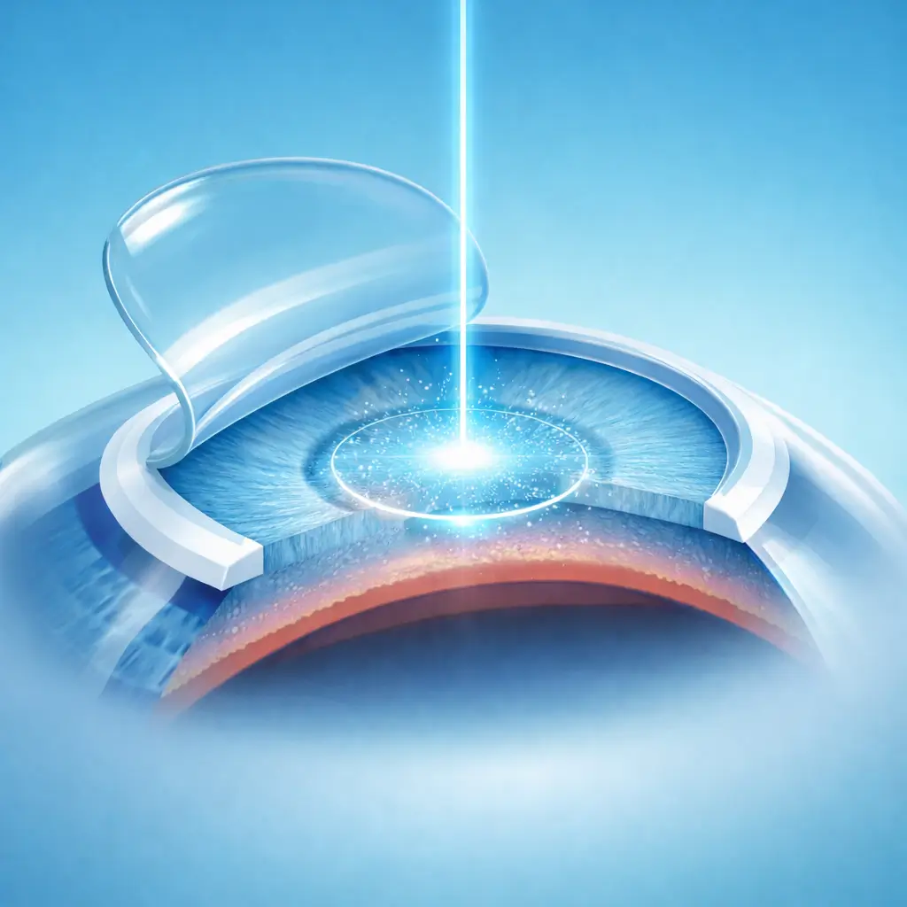
Femtosecond LASIK (FS-LASIK) combines femtosecond laser flap creation with excimer laser stromal ablation and represents the highest cumulative volume of any corneal refractive procedure worldwide. A femtosecond laser creates a uniform corneal flap of precisely defined thickness (typically 100-120 μm) with superior consistency compared with traditional microkeratome blades. The flap is reflected, and an excimer laser — following a personalized ablation profile that may be wavefront-guided or topography-guided — reshapes the stromal bed to correct the refractive error. The flap is then repositioned without sutures. FS-LASIK's key advantage is its wide prescription range: myopia up to approximately -15.00 D, hyperopia up to +6.00 D, and astigmatism up to 6.00 D. Vision recovers rapidly, often reaching serviceable acuity by the following morning. Principal limitations include flap-related risks (dislodgement, striae) and greater corneal nerve disruption than SMILE, resulting in a higher incidence of postoperative dry eye. Leading refractive centers in China's major cities operate multiple femtosecond platforms (Zeiss VisuMax, Alcon IntraLase/iFS) paired with advanced excimer systems (Alcon WaveLight EX500, Schwind Amaris, Zeiss MEL90) that enable individualized wavefront-optimized ablation profiles to enhance night-vision quality.
Quick Reference
Treatment
15 mins – 30 mins
Observation
1 hours – 2 hours
Est. Cost
$1,900 – $4,700
Department
Ophthalmology
Who Is This For
Is Femtosecond LASIK Laser Surgery (FS-LASIK) Right for You?
Good Candidates
- Age ≥18 with stable myopia for at least 2 years
- Wide prescription range: myopia up to ~-15.00 D, hyperopia up to +6.00 D, astigmatism up to 6.00 D
- Adequate corneal thickness (central pachymetry ≥500 μm; residual stromal bed ≥250 μm post-ablation)
- High astigmatism requiring correction beyond SMILE's range
- Normal corneal topography with no keratoconus features
May Not Be Suitable
- Keratoconus or suspicious corneal topography (contraindication to all flap-based procedures)
- Thin cornea insufficient to accommodate the flap and a safe residual stromal bed
- Significant dry eye disease (LASIK carries a higher risk of exacerbating dry eye than SMILE)
- Contact-sport athletes (boxing, combat sports) due to theoretical flap dislodgement risk with eye trauma
- Progressive myopia within the past 2 years
Step-by-Step Process
How Femtosecond LASIK Laser Surgery (FS-LASIK) Works
Preparation and Topical Anesthesia
The patient lies supine and receives topical anesthetic drops. The eye is sterilized and draped; a lid speculum is placed. Reference marks are placed on the cornea for accurate flap repositioning.
Femtosecond Flap Creation
A suction ring is applied; the femtosecond laser scans the predetermined stromal depth to create a flap ~8-9 mm in diameter and 100-120 μm thick, with a superior hinge of approximately 50°. Laser application takes about 15-20 seconds.
Flap Reflection
Once suction is released, a microhoe gently separates the flap from its edge and folds it back onto the hinge, exposing the stromal bed. Balanced salt solution keeps the exposed stroma moist.
Excimer Laser Ablation
The excimer laser delivers a personalized ablation profile (wavefront-guided or topography-guided) to the stromal bed. A high-speed eye-tracker continuously monitors eye position and adjusts the laser in real time. Ablation duration is approximately 20-60 seconds depending on prescription.
Flap Repositioning
The stromal bed is rinsed with balanced salt solution. The flap is carefully repositioned using reference marks for alignment and smoothed with a micro-spatula to eliminate any striae. Adhesion occurs without sutures via epithelial sealing and intraocular pressure.
Completion and Discharge
Antibiotic drops are instilled; a bandage contact lens may be placed to protect the flap edge. The patient rests in the recovery area for 60-120 minutes before discharge. Eyes-closed rest for the remainder of the day is strongly advised.
Both eyes are treated in a single visit. Follow-up visits are at day 1, week 1, months 1, 3, and 6, then annually.
Cost Information
Cost Estimate for Femtosecond LASIK Laser Surgery (FS-LASIK)
Estimated Price Range
$1,900 – $4,700
What's Included
Public tier-3A International Medical Department: approximately ¥13,000-22,000 (both eyes); premium private eye centers: approximately ¥20,000-32,000 (both eyes). Differentiated techniques such as Q-LASIK may carry slightly higher fees.
Before Your Visit
What to Prepare
Required Tests & Examinations
If you already have recent valid test results, bring the reports. If not, these assessments can usually be completed in China before the procedure.
Cycloplegic refraction (accurate refractive data including astigmatism axis)
Corneal topography + tomography (rule out keratoconus)
Corneal pachymetry (ensure safe residual stromal bed after planned flap and ablation)
Wavefront aberrometry (for personalized, wavefront-guided ablation profile)
Tear function evaluation (TBUT, Schirmer test, meibomian gland assessment)
Dilated fundus exam (rule out peripheral retinal degeneration, especially in high myopia)
Documents & Materials to Bring
Required to Bring
Refraction report obtained after discontinuing soft contact lenses for ≥1 week
Refraction records from the past 1-2 years confirming a stable prescription
Passport and valid visa
Records of any prior ocular conditions or surgeries (if applicable)
After Treatment
Recovery & Follow-Up
Rest with eyes closed on the day of surgery to allow early flap adhesion — avoid screens and reading for the first evening
Do not rub the eyes for at least 1 month; the flap remains vulnerable to dislodgement during the early healing phase
Avoid high-impact activities (contact sports, swimming) for at least 1 month
Use prescribed antibiotic + steroid drops for 2-4 weeks; preservative-free artificial tears for 3-6 months to manage dry eye
Postoperative dry eye is common and usually temporary; omega-3 supplementation can assist recovery
Night halos and starbursts are normal early findings and typically diminish as corneal nerves regenerate over 3-6 months
Follow-Up Schedule
Day-1 follow-up is mandatory to assess flap status and early visual acuity. Week-1 confirms flap adhesion. Months 1 and 3 verify the refractive outcome.
Related Conditions
Conditions This Procedure Treats
Ready to Plan Femtosecond LASIK Laser Surgery (FS-LASIK) in China?
Let Carevia help you find the right hospital, coordinate your treatment, and arrange every detail of your medical trip.
Frequently Asked Questions
Need personalized guidance?
Our care coordinators can help you assess whether this procedure fits your situation.
Contact Us