Apical Barrier Technique
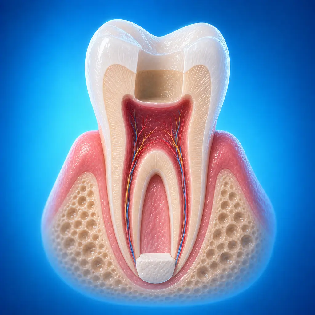
Bioceramic material is used to create an artificial barrier at an open apex, shortening the root canal treatment course for immature permanent teeth with incomplete apical closure.
The apical barrier technique uses MTA or other bioceramic material to create an artificial barrier in the apical area of necrotic immature permanent teeth with open apices, followed by root canal filling. Compared with traditional long-term apexification, it can shorten the treatment course and reduce the risk of root fracture during prolonged medication sealing. Indication selection, barrier thickness, quality of canal disinfection, and postoperative coronal sealing all affect prognosis, and regular imaging follow-up remains necessary.
Quick Reference
Treatment
1 hours – 2 hours
Observation
0 mins – 1 days
Est. Cost
$200 – $300
Department
Pediatric Dentistry
Who Is This For
Is Apical Barrier Technique Right for You?
Good Candidates
- Permanent teeth with pulp necrosis, periapical periodontitis, and an incompletely developed apex
- Permanent teeth that underwent long-term apexification but did not form an apical barrier
May Not Be Suitable
- Reversible pulpitis where pulp function can recover
- Infection limited to coronal pulp without root pulp involvement
- Root development less than 1/2 complete or extensive periapical bone destruction
- Permanent teeth with fully developed roots
Step-by-Step Process
How Apical Barrier Technique Works
Isolation and Access
Under rubber dam isolation, open the pulp chamber and confirm canal morphology, the extent of the open apex, and root canal wall thickness.
Canal Cleaning and Disinfection
Gently remove necrotic pulp and infected material, with irrigation and intracanal disinfection, while avoiding unnecessary weakening of thin root canal walls.
Apical Barrier Preparation
Place MTA or bioceramic material in the apical area to form a sealable artificial apical barrier, then temporarily seal and wait for the material to harden.
Root Canal Filling and Coronal Seal
After the barrier is stable, complete root canal filling and a tight coronal seal. Protective restoration may be needed to reduce root fracture risk.
Regular Imaging Follow-up
Follow up as instructed to assess periapical healing, barrier position, and root fracture risk. Return earlier if pain, swelling, or a sinus tract appears.
Usually completed in 2-3 visits: the first visit includes root canal preparation and disinfection plus barrier material placement; the second visit completes warm gutta-percha root canal filling and coronal restoration. Regular follow-up is needed afterward.
Cost Information
Cost Estimate for Apical Barrier Technique
Estimated Price Range
$200 – $300
What's Included
Fees usually include anesthesia, root canal disinfection, bioactive ceramic barrier material, microscope-assisted procedures, and later filling restoration. Final cost depends on material type and root canal complexity.
Before Your Visit
What to Prepare
Required Tests & Examinations
If you already have recent valid test results, bring the reports. If not, these assessments can usually be completed in China before the procedure.
X-ray to assess root development
Dental specialty examination and pulp vitality testing to determine pulp status
Detailed medical history
Documents & Materials to Bring
Required to Bring
Recent dental imaging, such as periapical radiograph or CBCT if available
Previous dental treatment records
Record of dental trauma, fractured dens evaginatus, or previous infection course if applicable
Medication allergy history
General medical history information
Companion & Support
An adult companion is recommended for postoperative pickup and observation. For general anesthesia, sedation, larger procedures, child patients, or patients with limited mobility, arrange accompaniment according to hospital requirements.
After Treatment
Recovery & Follow-Up
Mild soreness or swelling sensation may occur for 1-2 days after surgery and is normal.
Two hours after surgery, warm or cool liquid food may be eaten. Avoid chewing hard objects on the affected side for 2 weeks.
Maintain oral hygiene. For child patients, parents should supervise oral hygiene and use fluoride toothpaste to prevent secondary caries.
If clinical symptoms persist or worsen, return promptly to rule out infection.
Follow-Up Schedule
Usually every 3 months, focusing on periapical healing, barrier position, and coronal seal. Return earlier if pain, swelling, or a sinus tract occurs.
Related Conditions
Conditions This Procedure Treats
Ready to Plan Apical Barrier Technique in China?
Let Carevia help you find the right hospital, coordinate your treatment, and arrange every detail of your medical trip.
Frequently Asked Questions
Need personalized guidance?
Our care coordinators can help you assess whether this procedure fits your situation.
Contact Us