Sialolithiasis
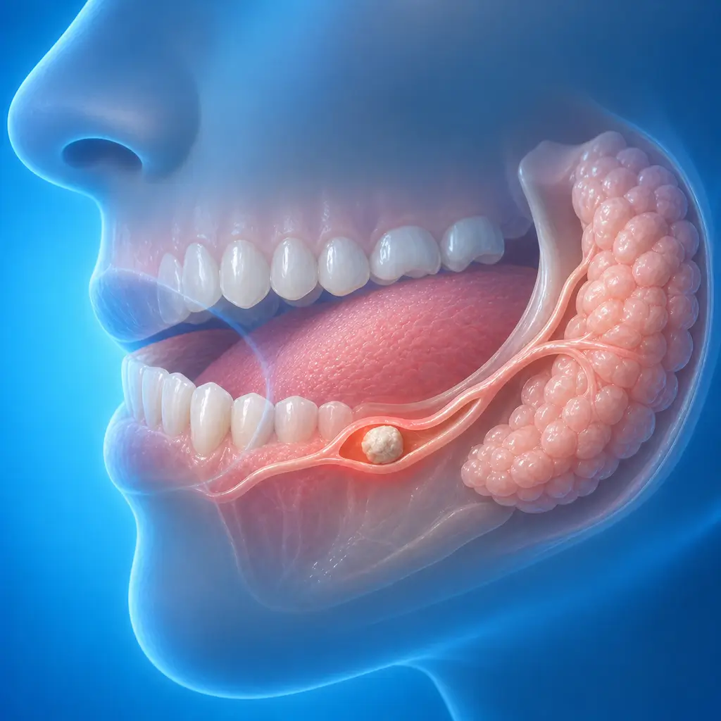
Sialolithiasis is a calcified mass within a salivary gland duct or gland that blocks saliva drainage, causing recurrent swelling and pain during meals. It is the most common salivary gland disease.
Common Symptoms
Recognizing Sialolithiasis
Focus on the most useful decision cues first: common symptoms, the patients or situations that usually prompt review, and any signs that need faster assessment.
Common Symptoms
Signs patients often notice before evaluation
Post-meal swelling: the gland rapidly enlarges and becomes distended and painful after eating, then gradually subsides 1-2 hours after meals. Palpation shows an enlarged, firm, tender gland. The duct opening is red and swollen, and pressing the gland may produce small amounts of pus or thick saliva
Secondary infection: persistent gland redness, swelling, heat, and pain with fever, headache, and fatigue. Pus drains from the duct opening, with limited mouth opening and swallowing difficulty
Late stage: gland fibrosis and atrophy. Post-meal swelling decreases, but the gland becomes hard and smaller
When to Seek Evaluation
Typical patients and situations that warrant review
Adults, slightly more often in men than women
Repeated swelling and pain in the submandibular or parotid area after eating, resolving 1-2 hours after meals
Persistent red, swollen, hot, painful gland with fever and pus from the duct opening
A hard stone can be felt at the intraoral duct opening
Radiographs, ultrasound, or CT show a salivary gland stone
Treatment Approaches
Treatment Directions for Sialolithiasis
Conservative treatment, minimally invasive stone removal, or gland removal is selected based on stone location, size, number, and gland function
Conservative treatment includes stimulating saliva, drinking more water, gland massage, and warm compresses. It is suitable for tiny or sandy stones
Intraoral duct incision and stone removal is suitable for palpable stones in the anterior or middle segment of the submandibular duct. Under local anesthesia, the duct wall is opened and the stone is removed while preserving the gland
Sialendoscopic stone removal is suitable for posterior duct stones, intraglandular stones, and multiple stones. Stones are grasped or retrieved with a basket under endoscopy, and large stones may be fragmented with laser assistance
Gland removal is suitable when deep intraglandular stones cannot be completely removed, the gland is atrophic, or infections recur. The gland and stones are completely removed
What usually shapes the treatment plan
Clinical Assessment
Key Assessments for Sialolithiasis
These are the main areas doctors usually review first. If you already have relevant test or imaging reports, bring them to speed up the assessment. They are helpful but not required, and the same workup can also be completed in China.
Stone location
Size
Number
Whether the gland is swollen
Tenderness
Texture
Whether the duct opening is red and swollen
Pus discharge
Frequency of post-meal swelling episodes
History of secondary infection
Before You Travel
How to Prepare
Bring previous systemic disease and treatment history, and recent imaging studies if available
Planning Notes
Pre-Assessment Required
An oral specialist should perform an intraoral examination and, as appropriate, periodontal probing, pulp vitality testing, periapical radiographs, panoramic radiographs, or CBCT before determining the treatment plan. Key checks include clinical examination with bimanual palpation along the duct, pressing the gland to observe saliva flow and pus; radiographs, such as floor-of-mouth occlusal film for radiopaque submandibular stones and parotid sialography for duct filling defects; CT to show exact stone location, size, number, and relationship to surrounding tissues to guide the surgical approach; and sialendoscopy entering through the duct opening to directly view stone location and size and perform endoscopic removal when possible. Bring clinical examination, CT, and sialendoscopy records if available.
Remote Pre-Assessment
Intraoral photos, the course of pain/swelling, previous dental records, and imaging can be submitted remotely for preliminary triage, urgency assessment, and an estimated treatment direction. Final diagnosis still requires in-person intraoral examination and necessary imaging.
Multidisciplinary Assessment
Medical History Important
Previous dental treatment history, imaging, allergy history, anticoagulant/bisphosphonate use, diabetes, and immune-related diseases can affect diagnosis, anesthesia, bleeding and infection risk, and treatment selection.
Ready to Explore Treatment for Sialolithiasis?
Let Carevia help you connect with the right specialists, compare hospitals, and plan your medical trip to China.