Alveolar bone hyperplasia
Alveolar bone hyperplasia is localized excessive growth of alveolar bone forming a bony prominence. It is usually a benign, asymptomatic anatomical variation, but surgical removal is needed when it interferes with denture restoration, implant placement, or causes mucosal trauma.
Common Symptoms
Recognizing Alveolar bone hyperplasia
Focus on the most useful decision cues first: common symptoms, the patients or situations that usually prompt review, and any signs that need faster assessment.
Common Symptoms
Signs patients often notice before evaluation
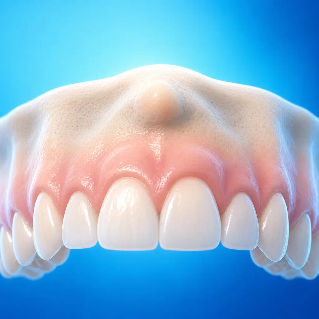
A bony prominence can be felt in the mouth
Interferes with denture wearing, causing rocking, instability, pressure pain, or ulcers
When to Seek Evaluation
Typical patients and situations that warrant review
More common in middle-aged and older adults
Patients after tooth extraction
A bony prominence can be felt in the mouth
Interferes with denture wearing, causing rocking, instability, pressure pain, or ulcers
Interferes with implant placement
A bone spicule forms after extraction and affects denture wearing
Treatment Approaches
Treatment Directions for Alveolar bone hyperplasia
Asymptomatic lesions that do not affect function can be observed conservatively
Symptomatic lesions or those affecting function are treated with alveoloplasty
What usually shapes the treatment plan
Clinical Assessment
Key Assessments for Alveolar bone hyperplasia
These are the main areas doctors usually review first. If you already have relevant test or imaging reports, bring them to speed up the assessment. They are helpful but not required, and the same workup can also be completed in China.
Location of the bony prominence
Size
Surface mucosal condition and tenderness
Whether it affects denture wearing
Whether it affects implant placement
Radiographs or CBCT to assess the extent of the bony prominence and relationship to nerves and blood vessels
Relationship to tooth roots, guiding surgical approach and depth
Before You Travel
How to Prepare
Bring dentures, recent imaging records, systemic disease history, and dental history
Planning Notes
Pre-Assessment Required
An oral specialist should perform an intraoral examination and, as appropriate, periodontal probing, pulp vitality testing, periapical radiographs, panoramic radiographs, or CBCT before determining the treatment plan. Key checks include specialist oral examination: visual assessment of location, size, shape, and surface mucosa of the bony prominence; palpation for texture, mobility, and tenderness; denture try-in to assess denture base fit, rocking, stability, and pressure pain; and CBCT to assess the extent of the prominence and its relationship to the mandibular canal, mental foramen, tooth roots, maxillary sinus, and other surrounding structures. Bring specialist examination and imaging records if available.
Remote Pre-Assessment
Intraoral photos, the course of pain/swelling, previous dental records, and imaging can be submitted remotely for preliminary triage, urgency assessment, and an estimated treatment direction. Final diagnosis still requires in-person intraoral examination and necessary imaging.
Multidisciplinary Assessment
Medical History Important
Previous dental treatment history, imaging, allergy history, anticoagulant/bisphosphonate use, diabetes, and immune-related diseases can affect diagnosis, anesthesia, bleeding and infection risk, and treatment selection.
Ready to Explore Treatment for Alveolar bone hyperplasia?
Let Carevia help you connect with the right specialists, compare hospitals, and plan your medical trip to China.