Dens invaginatus
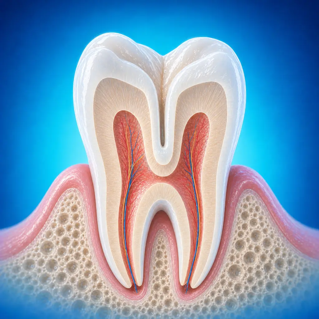
Dens invaginatus is a developmental abnormality of tooth shape in which enamel or dentin folds inward toward the pulp cavity, forming pouch-like structures of varying depth. It easily retains bacteria and can lead to early pulp infection and periapical disease.
Common Symptoms
Recognizing Dens invaginatus
Focus on the most useful decision cues first: common symptoms, the patients or situations that usually prompt review, and any signs that need faster assessment.
Common Symptoms
Signs patients often notice before evaluation
Mild dens invaginatus, such as an abnormal lingual pit, may cause no subjective symptoms and be found incidentally during oral examination
If the invagination is deep and bacterial accumulation causes pulpitis or periapical periodontitis, symptoms of pulpitis or periapical periodontitis may occur, such as pain and temperature sensitivity
When to Seek Evaluation
Typical patients and situations that warrant review
Common during the mixed dentition and immature permanent tooth stage
A deep pit, groove, or abnormal projection on the lingual side of a maxillary anterior tooth or premolar, with cold/heat sensitivity or spontaneous pain
Symptoms of pulpitis or periapical periodontitis without obvious caries in the affected tooth
Radiographs show a tooth-within-a-tooth or invaginated structure with a periapical radiolucency
Recurrent gum abscess corresponding to an abnormally shaped tooth
Treatment Approaches
Treatment Directions for Dens invaginatus
For shallow invagination without clinical symptoms, manage as deep caries by removing softened tissue and performing indirect pulp capping. If pulp exposure occurs during caries removal, direct pulp capping, pulpotomy, apexification, or apical barrier treatment is selected based on pulp status and root development
For deeper invagination in a vital tooth, gingival incision and flap reflection may be performed; shallow invaginations can be reshaped by grinding the pit and groove
For deeper invaginations, after cavity preparation, tooth filling restoration is performed, the wound is irrigated with saline and sutured, and a periodontal dressing is placed
For nonvital teeth, root canal treatment is followed by management of the pit and groove morphology
What usually shapes the treatment plan
Clinical Assessment
Key Assessments for Dens invaginatus
These are the main areas doctors usually review first. If you already have relevant test or imaging reports, bring them to speed up the assessment. They are helpful but not required, and the same workup can also be completed in China.
Type and depth of invagination
Pulp vitality status, normal, reversible, irreversible, or necrotic
Degree of root development, including whether the apical foramen is closed in immature permanent teeth
Whether periapical bone destruction is present and its extent
Whether the affected tooth can be retained
Before You Travel
How to Prepare
Bring imaging and dental treatment records
Maintain oral cleanliness and prepare the child psychologically in advance
Planning Notes
Pre-Assessment Required
An oral specialist should perform an intraoral examination and, as appropriate, periodontal probing, pulp vitality testing, periapical radiographs, panoramic radiographs, or CBCT before determining the treatment plan. Key checks include specialist oral examination: visual inspection of lingual morphology for a deep pit, groove, or abnormal projection; probing the fracture surface for a pulp exposure opening; percussion to assess periapical inflammation; and checking for gingival redness, swelling, or sinus tract. Pulp vitality testing is used to determine whether the pulp is normal, sensitive, or necrotic. Bring imaging and history records if available.
Remote Pre-Assessment
Intraoral photos, the course of pain/swelling, previous dental records, and imaging can be submitted remotely for preliminary triage, urgency assessment, and an estimated treatment direction. Final diagnosis still requires in-person intraoral examination and necessary imaging.
Multidisciplinary Assessment
Depending on the condition, joint evaluation by oral and maxillofacial surgery, endodontics, periodontics, prosthodontics, orthodontics, imaging, anesthesia, or other related disciplines is recommended, especially for complex infection, tumors, trauma, jaw lesions, or high systemic disease risk.
Medical History Important
Previous dental treatment history, imaging, allergy history, anticoagulant/bisphosphonate use, diabetes, and immune-related diseases can affect diagnosis, anesthesia, bleeding and infection risk, and treatment selection.
Ready to Explore Treatment for Dens invaginatus?
Let Carevia help you connect with the right specialists, compare hospitals, and plan your medical trip to China.