Central cusp deformity
Central cusp deformity is a developmental tooth abnormality presenting as an extra cusp in the center of the occlusal surface of a premolar, especially the mandibular second premolar. It can fracture or wear during chewing, exposing the pulp and causing pulpitis and periapical disease.
Common Symptoms
Recognizing Central cusp deformity
Focus on the most useful decision cues first: common symptoms, the patients or situations that usually prompt review, and any signs that need faster assessment.
Common Symptoms
Signs patients often notice before evaluation
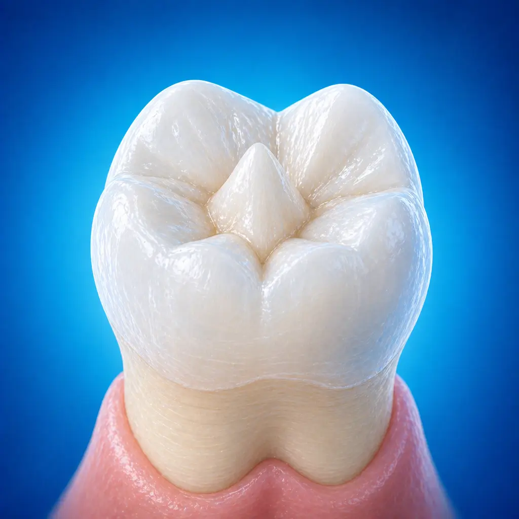
Abnormal cusp shape: a conical or rounded extra cusp is visible in the center of the tooth's occlusal surface
If the cusp is intact and the pulp is not exposed, there are usually no subjective symptoms
When cusp wear or fracture exposes dentin, pulpitis symptoms may occur, such as transient sensitivity to cold or heat, spontaneous pain, or night pain. After pulp necrosis, the crown may gradually turn gray or dark
When to Seek Evaluation
Typical patients and situations that warrant review
Common during the mixed dentition and immature permanent tooth period, about ages 7-14
A conical extra cusp on the occlusal surface with cold/heat sensitivity or spontaneous pain
The cusp has fractured, with visible pulp exposure or a pinpoint pulp exposure opening
Radiographs show a periapical radiolucency, and the affected tooth has no obvious caries or trauma history
Urgent Assessment
If a central cusp deformity fractures and severe spontaneous pain, night pain, biting pain, gingival swelling, or a sinus tract develops, seek prompt dental evaluation of the pulp and periapical area. Delayed treatment in immature permanent teeth may affect continued root development.
Treatment Approaches
Treatment Directions for Central cusp deformity
Rounded cusps that do not interfere may be left untreated
For sharp and long cusps, one-stage grinding under local anesthesia and disinfection followed by cavity preparation and pulp capping may be performed; alternatively, small repeated adjustments may be used so the pulp horn forms enough reparative dentin to avoid exposure
If the cusp has fractured and exposed the pulp but pulp vitality is normal, direct pulp capping or pulpotomy may be performed to preserve vital pulp and promote continued root development
If the pulp is necrotic but root development is incomplete, apexification or an apical barrier procedure is performed
What usually shapes the treatment plan
Clinical Assessment
Key Assessments for Central cusp deformity
These are the main areas doctors usually review first. If you already have relevant test or imaging reports, bring them to speed up the assessment. They are helpful but not required, and the same workup can also be completed in China.
Confirm whether the affected tooth has a central cusp deformity and whether the cusp is intact
Whether pulp exposure is present and pulp vitality status
Whether there are clinical signs of pulpitis or periapical periodontitis
Degree of root development, including whether the apical foramen is closed
Whether periapical bone destruction is present and its extent
Whether the affected tooth can be retained
Child age and cooperation
Before You Travel
How to Prepare
Bring imaging and dental treatment records
Maintain oral cleanliness and prepare the child psychologically in advance
Planning Notes
Pre-Assessment Required
An oral specialist should perform an intraoral examination and, as appropriate, periodontal probing, pulp vitality testing, periapical radiographs, panoramic radiographs, or CBCT before determining the treatment plan. Key checks include visual examination of the premolar occlusal surface for extra cusps and whether the cusp is intact, worn, or fractured; probing the fracture surface for a pulp exposure opening; percussion to assess periapical inflammation; and checking for gingival redness, swelling, or sinus tract. Bring imaging and history records if available.
Remote Pre-Assessment
Intraoral photos, the course of pain/swelling, previous dental records, and imaging can be submitted remotely for preliminary triage, urgency assessment, and an estimated treatment direction. Final diagnosis still requires in-person intraoral examination and necessary imaging.
Multidisciplinary Assessment
Medical History Important
Previous dental treatment history, imaging, allergy history, anticoagulant/bisphosphonate use, diabetes, and immune-related diseases can affect diagnosis, anesthesia, bleeding and infection risk, and treatment selection.
Ready to Explore Treatment for Central cusp deformity?
Let Carevia help you connect with the right specialists, compare hospitals, and plan your medical trip to China.